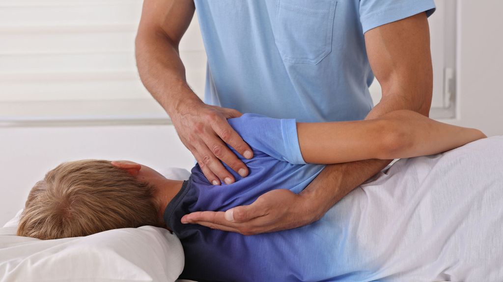
Adolescent Idiopathic Scoliosis
More than 1100 participants from all over the world joined the third online session of “Spine to go” – a free, nonprofit event offering further training and continuing education for physicians and therapists. International experts presented the history and pathophysiology of adolescent idiopathic scoliosis (AIS) and discussed treatment options like physiotherapy and bracing as well as surgical approaches.
Scoliosis is a three-dimensional deformity caused by complex tissue alterations changing the position and orientation of the vertebra in space. Idiopathic scoliosis affects 2–3% of the population. The adolescent form is the most common one which primarily occurs in females – especially the more severe curves that need surgical treatment. The etiology and initiation of scoliosis remain unclear. “We know that more than 90% of monozygotic twins and more than 60% of dizygotic twins demonstrate concordance regarding their idiopathic scoliosis,” explains Prof. Stefan Parent, MD, PhD, Department of Surgery, University of Montréal (Canada). Genetics like POC5 mutations or hormones like melatonin might contribute to the pathogenesis of AIS but most of the theories discuss abnormal biomechanical forces as the reason for developing AIS. “Smith et al. and later Dickson have proposed a buckling theory of spinal deformity. It’s based on a posterior tether causing hypokyphosis and progressive lordosis leading to a rotatory deformity and a lateral bending moment,” explains Parent. Bone growth influences scoliosis. Vertebrae have a growing cartilage at the junction between disc and vertebral body. The ring apophysis appears during growth and progressively ossifies until it unites with the body at the end of growth. “Stokes et al. described the vicious cycle of scoliosis. Initially you have some form of asymmetry which leads to wedging of vertebrae and discs. The spinal curvature increases and produces more asymmetric loading and asymmetric growth following the Hueter-Volkman law resulting in progressive wedging,” says Parent. According to Hueter-Volkman law, bone growth is retarded by increased mechanical compression and is accelerated by reduced loading. “Which curves will progress? Which curves are not likely to progress? These are very important questions to ask,” recommends Parent. Curves at risk for progression should be detected early. Known factors for progression include age, sex, initial Cobb angle, peak growth velocity and family history. Age respectively skeletal maturity is a significant factor determined by triradiate cartilage, Risser sign, Sanders score and menarchal status. The risk of curve progression is higher if patients present at a young age with a severe curve size. “We developed a model of the final Cobb angle in patients with AIS assessing initial 3D parameters: angle of the plane of maximal curvature, 3D wedging of two specific disc levels and apical intervertebral rotation. This model improved our ability to determine which patients are likely to progress,” states Parent.
Achieving self-correction with PSSE
“We are currently evaluating the effectiveness of therapeutic exercise in treating patients with AIS comparing Generic Therapeutic Exercise (GTE) and Physiotherapeutic Scoliosis Specific Exercise (PSSE),” explains Prof. Stefano Negrini, MD, Physical and Rehabilitation Medicine, University “La Statale”, Milan (Italy). “We found that PSSE reduced the asymmetry of the waist and improved the patient’s quality of life compared to no treatment. PSSE led to a greater reduction of the Cobb angle compared to GTE,” says Negrini. Exercises can reduce progression, avoid bracing and train functions to counteract the pathological conditions. They can be proposed alone or in combination with bracing to enhance the results of bracing. “Exercise as a stand-alone therapy is not indicated for curves above 30° Cobb as the risk for progression is too high,” explains Negrini. Exercise cannot achieve a correction of the spine. Instead, it reduces the postural collapse and educates the patient to keep a better posture against their scoliosis. “We act on Stoke’s vicious cycle aiming to reduce the asymmetrical load and growth with improved posture and movement,” states Negrini.
The main element of PSSE is a 3-dimensional self-correction. The patient is taught how to biomechanically achieve and keep a better posture. The correction is either trained through repetition or challenge. There are two major schools – the Schroth school from Germany and the Scientific Exercises Approach to Scoliosis (SEAS) from Italy. The Schroth therapy includes only one or two exercises that are learned intensely and repeated until perfection. In contrast, treatment with SEAS is based on performing thousands of different exercises that challenge the patient’s capacity to keep the self-corrected posture. “There is a progressive increase of the challenge to allow the patient to learn how to better correct the posture,” explains Negrini. “To put it in a nutshell: the Schroth school aims for the greatest possible self-correction through repetition whereas the SEAS focuses on the best achievable and keepable self-correction through challenge.”
Bracing works
Bracing aims to prevent a curve from getting worse during rapid growth but bracing is not a benign therapy – psychosocial problems occur and compliance is an issue. “When I started looking into this topic, I realized that bracing has actually never been subjected to a rigorous evaluation of its efficacy or effectiveness,” says Stuart L. Weinstein, MD, Orthopaedic Surgery, University of Iowa Hospitals and Clinics (USA). “We found inconclusive results whether braces worked to prevent the need for surgery. That’s why we conducted the bracing in AIS trial1,” states Weinstein. The trial investigated if braces lower the risk of curve progression to a surgical threshold in high-risk patients with AIS relative to observation only. A secondary aim was to determine the relationship between bracing time and curve response. “Our trial had to be stopped early for efficacy because bracing did help to prevent the need for surgery,” explains Weinstein. Furthermore, a significant association between increasing hours of brace wearing and treatment success was found. “If the brace was worn more than 13 hours a day there was a 90% chance of avoiding a curve to enter the surgical range,” says Weinstein. Further study showed that the use of the Risser sign as a marker for maturity was contributing to overtreatment of patients. To avoid overtreatment, a prognostic calculator2 was developed. It’s free to use and calculates a probability of a curve reaching the surgical threshold based on curve pattern, Cobb angle and Sanders score. The Sanders score correlates with peak growth velocity and needs to be assessed correctly. “We are currently working on a new calculator3 which will also be free to use to enhance shared decision making, to minimize overtreatment and to find a personalized bracing time,” finishes Weinstein.
First choice: posterior spinal fusion
The gold standard for surgical treatment of AIS is posterior spinal instrumentation and fusion (PSIF). Its goal is a 3-dimensional correction with a safe and solid fusion as short as possible to achieve a balanced spine and trunk. One of the most challenging but important parts is the preoperative planning. “We use the Lenke classification as a template to determine which curves to fuse. It recommends fusing the structural curves,” explains Prof. Ahmet Alanay, MD, Department of Orthopaedics and Traumatology, Acibadem Mehmet Ali Aydinlar University School of Medicine, Istanbul (Turkey). Clinical and radiographic assessments help to select the fusion levels during preoperative planning. Coronal and sagittal radiographs are evaluated to identify the first vertebra intersected by CSVL (central sacral vertical line) which can be accounted as the stable vertebra for selection of LIV (lowest instrumented vertebra). “Flexibility radiographs may help us to find the stable vertebra-to-be. By definition, it’s located above the stable vertebra, not bisected by CSVL in a standing radiograph but bisected or intersected by CSVL in the concave bending radiograph. We usually want our stable vertebra-to-be become parallel to S1,” states Alanay. Planning of sagittal alignment and kyphosis are equally important.
The main forces to correct a deformity include translation, rotation and Cantilever reduction. Multiple anchorage points are essential when performing PSIF. Surgeons need to have great expertise in order to choose the right maneuver for correction according to the specific case. “My preferred method is the following: Translation by using traction towers, pulling the apex with towel clips to help restoration of kyphosis, in situ bending, Cantilever correction via convex rods, segmental derotation, completed by compression and distraction,” explains Alanay.
Balanced spine and short fusion length with anterior correction
“Anterior scoliosis correction was very popular in the 1990s but it has lost popularity because it is technically demanding and young surgeons are no longer trained in these approaches”, says Prof. Ulf Liljenqvist, MD, Department of Spine Surgery and Scoliosis Center, St. Franziskus Hospital, Münster (Germany). But this technique provides essential advantages. “With anterior scoliosis correction you can reach certain goals that could not be reached with posterior techniques. Due to the segmental disc resection, you can achieve a superior derotation and ribhump correction,” states Liljenqvist. In most cases the fusion length is shorter compared to posterior techniques. The risk of infection or paraplegia is insignificant. “You also leave the back muscles untouched which is important for these young patients to regain their sporting activities quickly,” adds Liljenqvist. The main disadvantages are approach-related complications such as pleural effusion, lung volume restriction or postsympathectomy syndrome. The hospital stay is approximately two days longer and the range of available implant systems is limited. Anterior correction is indicated for Lenke type 1 curves – especially those which are hypokyphotic with a severe ribhump – and Lenke type 5 curves. Anterior and posterior techniques have comparable outcomes regarding the frontal plane. “Anterior correction can restore thoracic and rotational kyphosis in the sagittal plane and it enables derotation of the thoracic and lumbar spine in the transverse plane. Ribhump correction can be achieved by true segmental derotation,” notes Liljenqvist.
No more need for spinal fusion?
Vertebral tethering gains more popularity from year to year and according to Peter O. Newton, MD, Division of Orthopaedics and Scoliosis, Rady Children’s Hospital, San Diego (USA), it has a game-changing potential. “The great hope of anterior tethering is that we can maintain motion, modulate growth through the Hueter-Volkmann principles, truly reshape vertebrae in all three dimensions and finally achieve a lasting correction without performing a spinal fusion.” This technique is largely performed endoscopically with a thoracoscope. “Growth is what powers the correction. That’s why this is a technique for young patients who have a lot of growth left,” says Newton. The correction ranges widely and in general, the rate of correction is related to height velocity and Sanders score. “For Sanders 2 patients we see about 2,5 degrees correction per segment per year for about 2,5 years,” states Newton. Initial results are better compared to PSIF but after two years a rebound with loss of correction occurs. The rate of complications and revisions is still higher and the rate of cord failure increases over time. Further research is needed to solve these issues. “We are still in the very early generations of these implants. I remain committed to this concept because I do believe that there is a method and mechanism resulting in a lasting solution that doesn’t involve fusion which will ultimately be better for patients in the long haul,” says Newton.
Source:
Online course “Spine to go”, 19th February 2022
Literature:
1 Weinstein SL et al.: Effects of bracing in adolescents with idiopathic scoliosis. N Engl J Med 2013; 369(16): 1512-21 2 https://uichildrens.org/ais-prognosis-calculator-simplified 3 http://iihr-vl01.iihr.uiowa.edu/dev/bekir/braist_calc/calc.html
Das könnte Sie auch interessieren:

«Die Hüfte war damals ein kaum verstandenes Gelenk»
Im Gespräch mit Leading Opinions Rheumatologie & Orthopädie erzählt Prof. Dr. med. Michael Dienst, München, nach seinem Vortrag am Hip-Symposium Bern 2026 von der Entwicklung der ...
Die Schulter im Sport
Die Schulter ermöglicht wie kein anderes Gelenk die Kombination aus maximaler Beweglichkeit und funktioneller Präzision – und ist gerade deshalb im Sport besonders verletzungsanfällig. ...
Handverletzungen im Sport
Im modernen Breitensport ist ein stetig steigender Aktivitätstrend zuverzeichnen, der jedoch mit einer Zunahme spezifischer Verletzungsmuster einhergeht.So entfallen mittlerweile bis zu ...